In the realm of heart care, 2D echocardiography stands out as a vital tool for diagnosing and managing various cardiovascular conditions. As one of the best heart care hospitals in the region, we are proud to offer cutting-edge echocardiography services. Our team of experts, including the best cardiologist in Rajnandgaon, utilizes this technology to provide precise assessments and tailored treatment plans.
What is 2D Echocardiography?
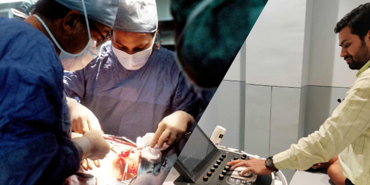
2D echocardiography, or two-dimensional echocardiography, is a non-invasive imaging technique that uses ultrasound waves to create real-time images of the heart. This method helps visualize the heart's structure and function, allowing cardiologists to diagnose and monitor heart conditions accurately. The procedure involves placing a transducer on the skin, which emits high-frequency sound waves that bounce off the heart structures and are then converted into images on a monitor.
Benefits of 2D Echocardiography
2D echocardiography offers several benefits:
- Non-Invasive: It doesn’t require any incisions or injections.
- Real-Time Imaging: Provides live images of the heart’s movement.
- Detailed Visualization: Allows for detailed examination of heart chambers, valves, and blood flow.
- Risk-Free: It uses sound waves rather than radiation, making it safe for repeated use.
2D Echocardiography for Adults
Common Indications
For adults, 2D echocardiography is often used to diagnose and evaluate:
- Heart Valve Disorders: Detecting conditions such as valve stenosis or regurgitation.
- Heart Failure: Assessing the heart's pumping ability and structure.
- Cardiomyopathy: Evaluating heart muscle abnormalities.
- Congenital Heart Defects: Identifying structural problems present from birth.
Procedure
The procedure for adults typically involves the following steps:
- Preparation: Patients may be asked to change into a hospital gown and lie down on an examination table.
- Ultrasound Gel Application: A gel is applied to the chest area to ensure good contact between the skin and the transducer.
- Imaging: The cardiologist moves the transducer over the chest to capture various angles of the heart.
- Analysis: The images are analyzed to assess the heart's size, shape, and function.
Post-Procedure
After the procedure, patients can resume their normal activities. The cardiologist will review the images and discuss the findings, providing recommendations for further treatment if necessary.
2D Echocardiography for Children
Special Considerations
In pediatric patients, 2D echocardiography plays a crucial role in:
- Congenital Heart Defects: Identifying and assessing heart defects present from birth.
- Acquired Heart Conditions: Evaluating conditions that develop over time, such as rheumatic fever.
- Pre-Surgical Planning: Providing detailed imaging to guide surgical or interventional procedures.
Procedure
The procedure for children involves similar steps as for adults but with added considerations for comfort:
- Preparation: Children may need to be calm and still, so sometimes sedation or distraction techniques are used.
- Ultrasound Gel Application: Gel is applied, and the transducer is used to capture images.
- Imaging: Special care is taken to ensure the procedure is as quick and comfortable as possible.
- Analysis: Images are reviewed to assess the child's heart condition.
Post-Procedure
Children generally recover quickly from the procedure. The cardiologist will discuss the results with the parents and outline any necessary follow-up steps.
2D Echocardiography for Fetuses
Importance of Fetal Echocardiography
Fetal echocardiography is a specialized application of 2D echocardiography used during pregnancy to assess the unborn baby's heart. It is particularly important for:
- Detecting Congenital Heart Defects: Identifying issues with the heart’s structure before birth.
- Monitoring Heart Function: Assessing the heart’s development and function throughout pregnancy.
- Planning for Delivery: Providing critical information for planning delivery and postnatal care.
Procedure
The procedure for fetal echocardiography includes:
- Preparation: The mother is positioned comfortably, and gel is applied to the abdomen.
- Ultrasound Imaging: The transducer is moved over the abdomen to capture images of the fetus's heart.
- Analysis: Detailed images are analyzed to assess the heart’s structure and function.
Post-Procedure
The results of a fetal echocardiogram are typically discussed with the parents, and any necessary follow-up or additional care plans are outlined.
Why Choose Us?
At our hospital, we pride ourselves on being a leader in heart care. With the expertise of the best cardiologist in Kawardha, we ensure that every patient receives top-notch care tailored to their specific needs. Our team’s dedication to using advanced diagnostic tools like 2D echocardiography allows us to provide accurate and effective treatment plans.
Conclusion
2D echocardiography is a powerful tool in modern cardiology, providing essential insights into heart health across all age groups. From adults to children and even fetuses, this technology helps in diagnosing, monitoring, and managing various heart conditions. Our hospital’s commitment to offering the highest level of care is reflected in our use of advanced echocardiography techniques and the expertise of our cardiologists. If you or your loved ones need a thorough heart evaluation, our team, including the best cardiologists in Rajnandgaon and Kawardha, is here to provide exceptional care and support.
Feel free to contact us for more information or to schedule an appointment. Our team is dedicated to ensuring your heart health is in the best possible hands.